Fact-sheet: Echinococcosis - Hydatid cyst
Updated on 03/23/2022 at 10:23 AM
View all RADEOS cases associated with this fact-sheet
Definition
Hepatic parasitic disease due to accidental ingestion by humans of the larval form of Echinococcus granulosus, originating from the dog (definitive host).
Other locations (brain, muscle, etc.) are possible.
Clinical features
Long clinical latency
Right upper quadrant pain
Abdominal mass
Compressive complications (vascular, biliary)
Rupture into the biliary tract, peritoneum, pleura, or lung
Ultrasound
Cystic lesion whose appearance depends on the lesion stage (see below).
The endocystic membranes are ribbon-like and hyperechoic.
CT
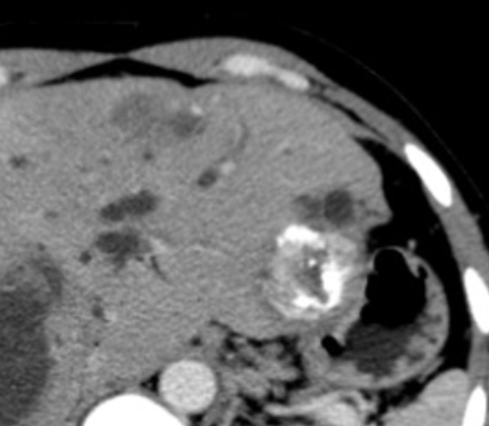
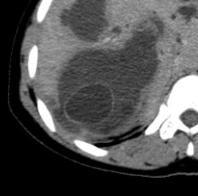
Cystic lesion with dense content (hydatid sludge), possibly harboring serpiginous internal membranes (snake sign, pathognomonic), +/- peripherally calcified. The appearance depends on the stage (see below)
MRI
On MRI, the endocystic membranes are hypointense on all sequences.
Classification
WHO classification.
Active forms
- Stage CE1: Anechoic cyst or one with faint dependent debris, with a relatively thick wall
- Stage CE2: Multilocular cyst with daughter vesicles
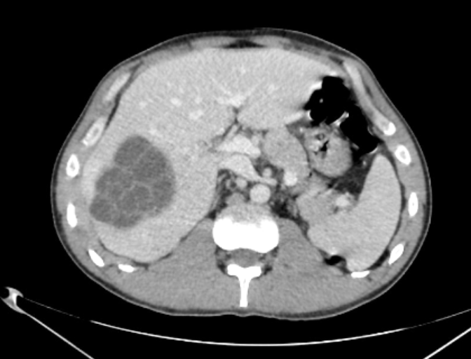
- Stage CE3, transitional stage suggesting a degenerating cyst = CE3A, fluid collection containing detached membranes; CE3B, solid matrix harboring daughter vesicles
Inactive forms corresponding to degenerated cysts
- Stage CE4: Heterogeneous, pseudotumoral solid matrix, without daughter vesicles
- Stage CE5: Calcified