
Fact-sheet: Cavernous angioma of brain
- Cerebral cavernoma
Updated on 10/11/2022 at 11:02 AM
View all RADEOS cases associated with this fact-sheet
Definition
Mulberry-shaped vascular malformation consisting of blood-filled cavities lined by a single layer of endothelial cells without interposed brain tissue (different from capillary telangiectasia). Macroscopy: mulberry-like, red-brown, of variable size, +/- calcific deposits. Location: -Supratentorial 80% - subcortical - temporal lobe. -Infratentorial 15% - pons. -Spinal cord and rare locations. Epidemiology: Prevalence of 0.5%. Sex ratio approximately 1. -Sporadic form: 80% -Familial form: autosomal dominant transmission - multiple locations. -Radiation-induced form: outside areas of maximal irradiation. Hemorrhagic risks ++: Female sex, prior hemorrhage, brainstem location.
Clinical features
- Asymptomatic, incidental finding ++.
- Headache (without hemorrhage)
- Epilepsy (cortico-subcortical location).
- Focal neurological signs.
CT
Mildly hyperdense lesion with irregular margins.
Sometimes calcified, rarely cystic.
No perilesional edema but sometimes perilesional hypodensity reflecting gliosis.
Possible mild enhancement after contrast injection
MRI
Cluster of loculi of variable signal intensity surrounded by a hemosiderin rim on T1/T2.
Ex: Subacute hemorrhage (hyperintense on T1 and T2) / chronic (hypointense on T1 and T2, more conspicuous on T2 GRE)
Frequent association with a developmental venous anomaly.
Hemosiderin rim accentuated on T2 GRE (blooming artifact).
Perilesional T1-hyperintense rim favoring cavernoma over hemorrhagic metastasis.
Mild enhancement.
Absence of visible arteriovenous shunt on MR angiography (differential diagnosis: AVM)
Vascular intervention
Little clinical value.
Lesion usually not visible, or slight blush on delayed images.
Frequent association with a developmental venous anomaly.
Management
- Follow-up – asymptomatic.
- Symptomatic treatment (epilepsy).
- Surgical resection: drug-resistant epilepsy. Hemorrhagic form. Accessible location.
- Radiosurgery: Inoperable forms with high hemorrhagic risk (brainstem)
Classification
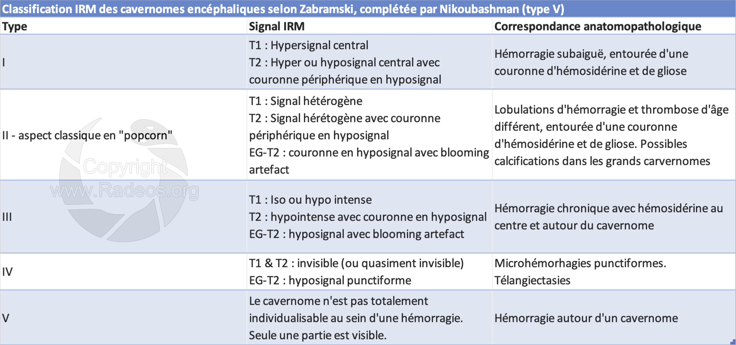
Zabramski classification
Differential diagnosis
-Melanoma metastasis: spontaneous T1 hyperintensity - melanin
-Hemorrhagic metastasis: perilesional T1-hyperintense rim favoring cavernoma
-AVM: intense serpiginous enhancement, high-flow arteriovenous shunt (MRA)